Identify the examination
Know what the technology measures and which clinical question it can help answer before reading any map or index.
Connect image quality, maps, parameters, and clinical context with a visual system for Pentacam, OCT, visual fields, biometry, and more — so you know what to check first and how the findings fit together.
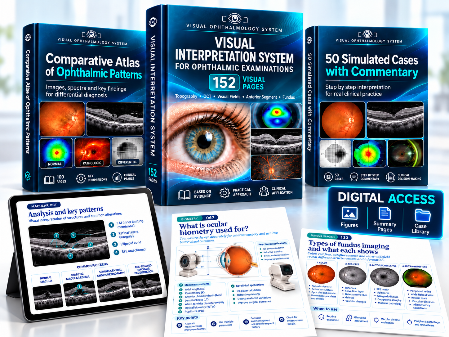
Turn the pages to preview selected topics and see how the system turns complex exam screens into a clear reading sequence.
Preview of the main resource with sample pages. The full system contains 152 pages across 13 modules.
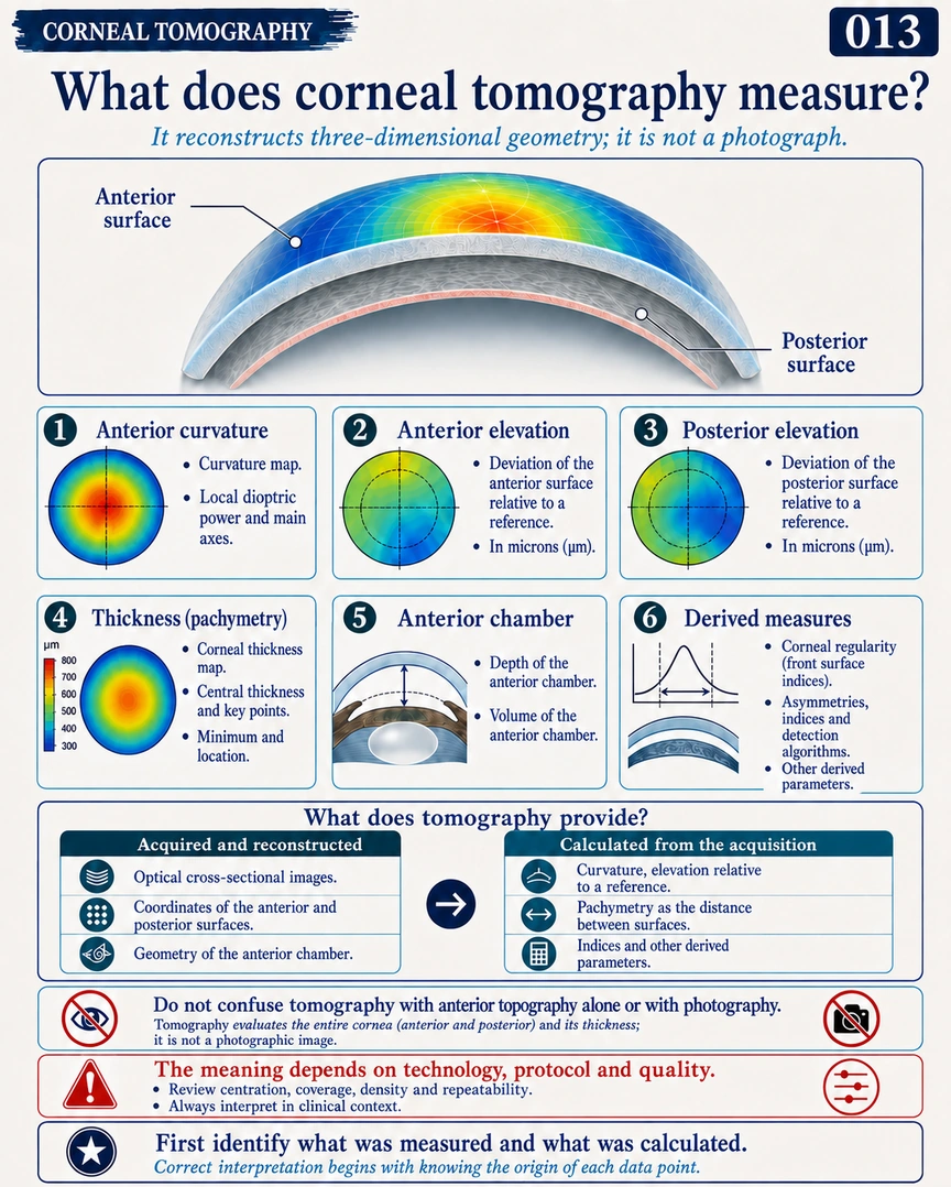
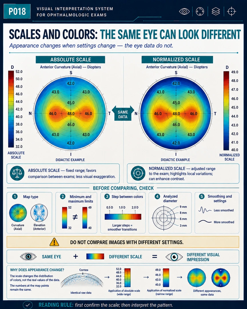
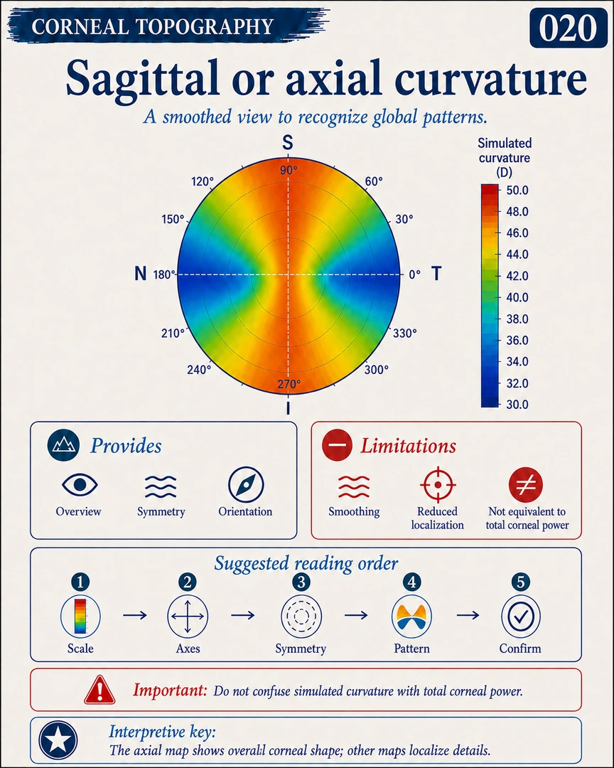
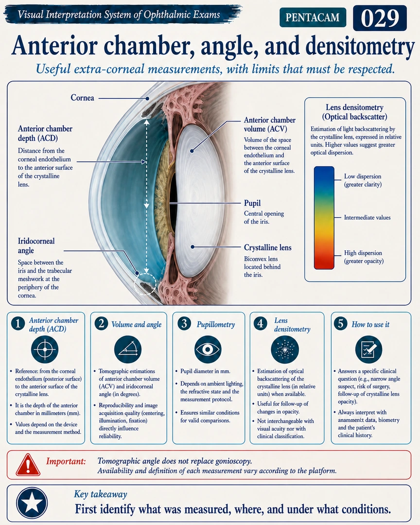
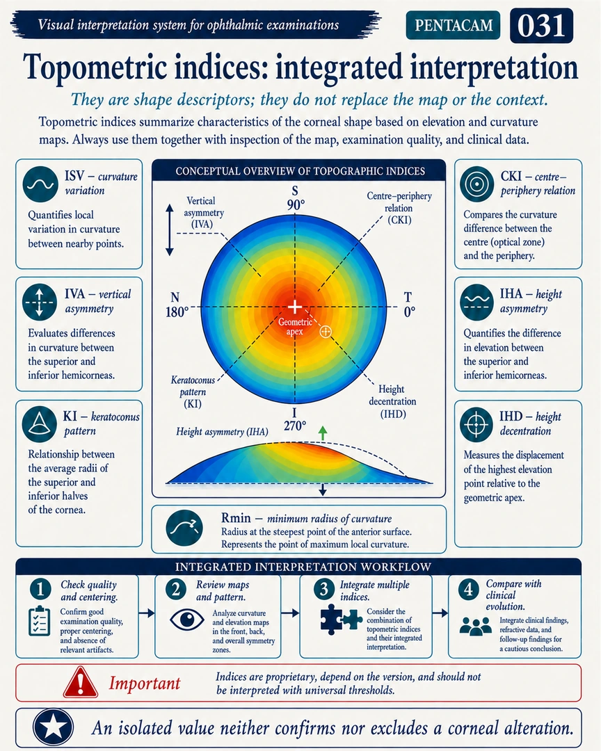
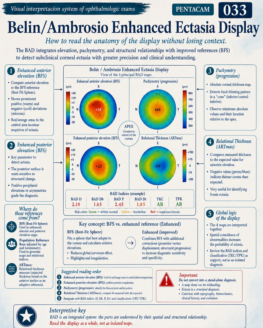
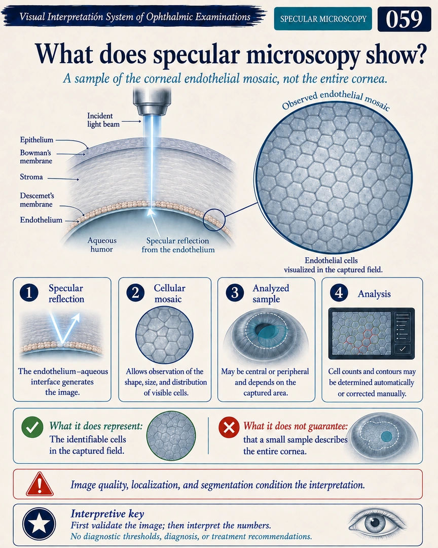
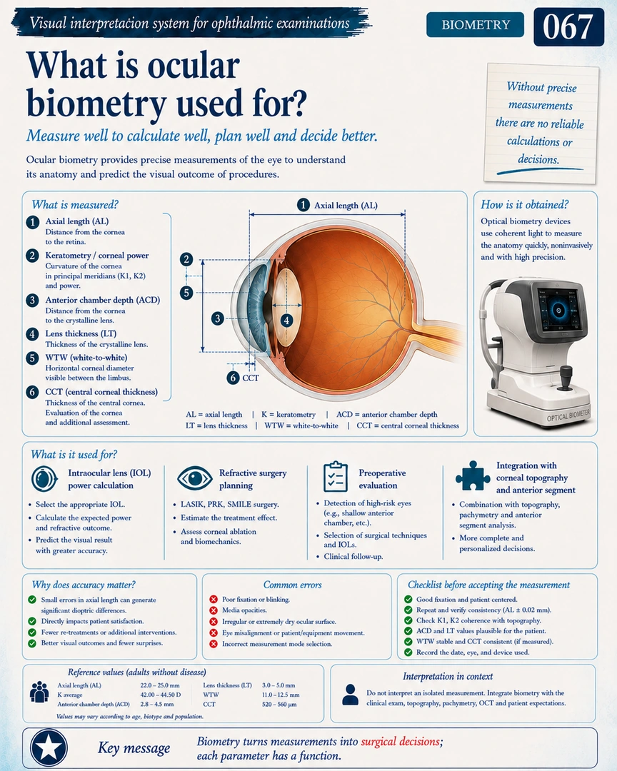
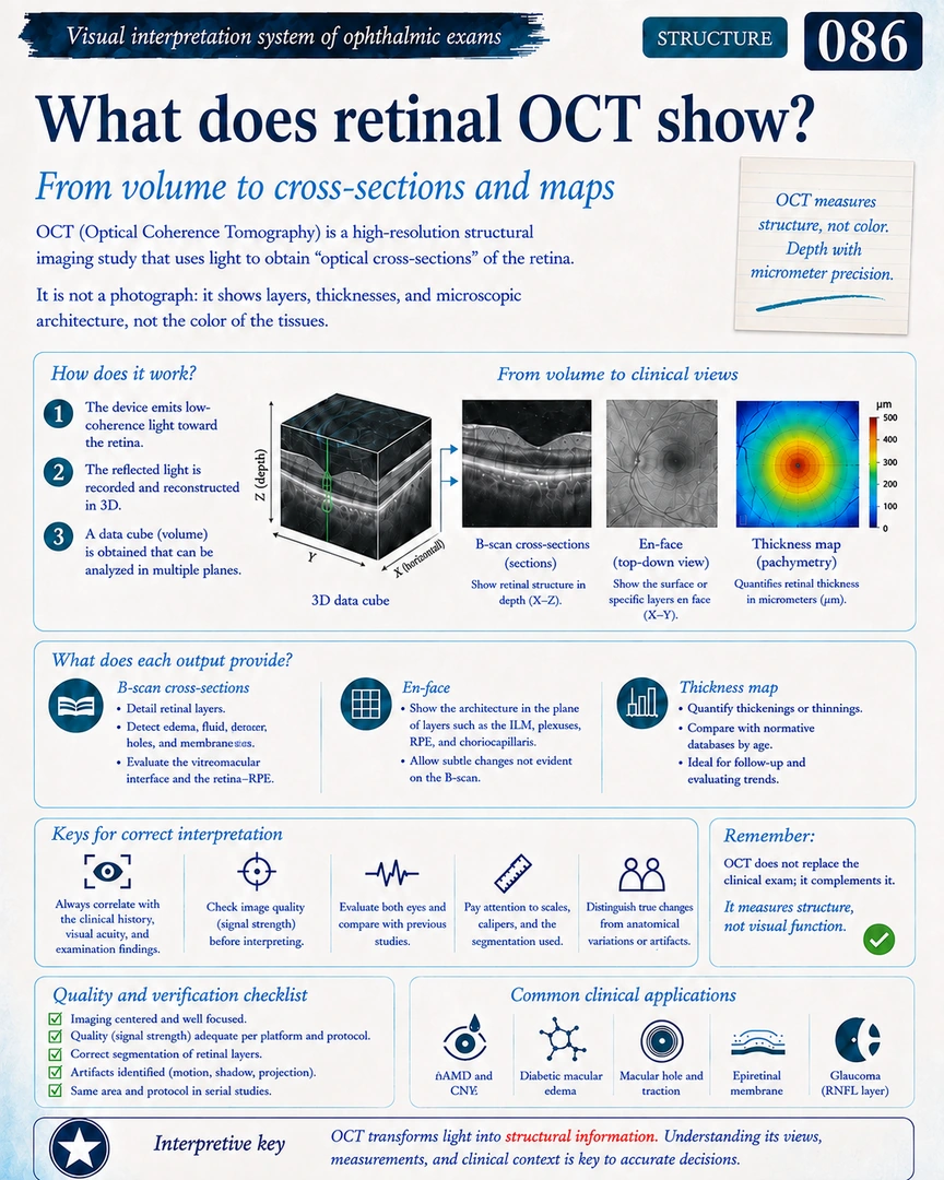
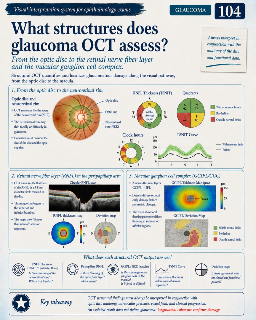
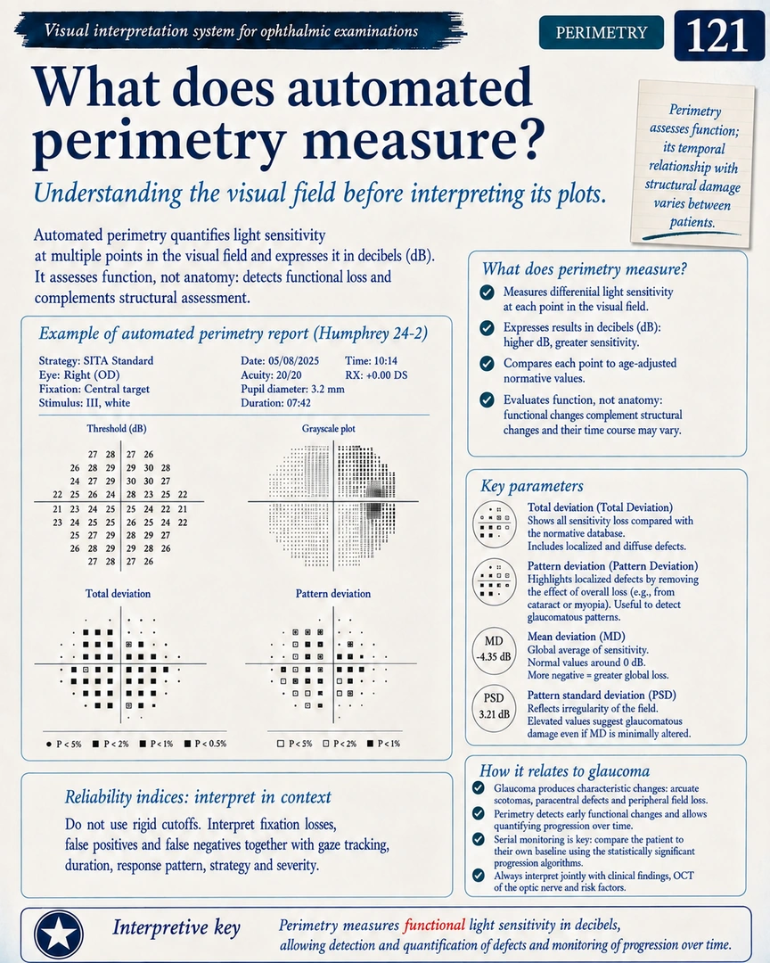
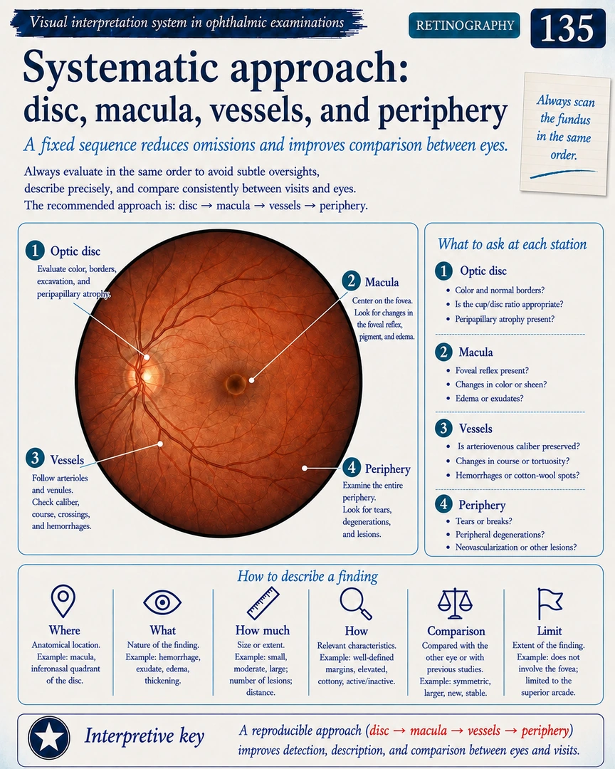
A visual structure that takes you from image quality and key parameters to pattern recognition and integrated case interpretation.
Know what the technology measures and which clinical question it can help answer before reading any map or index.
Check centration, acquisition, signal, and reliability before allowing a map or measurement to guide your interpretation.
Find the maps, curves, sectors, and indices that matter first without getting lost in secondary data.
Understand what each value represents and how it should be read within the context of the complete examination.
Compare normal, suspicious, and pathological findings side by side to make the differences easier to recognize.
Bring image quality, maps, parameters, and clinical context together to practice a more structured interpretation.
A visual library for students, trainees, and professionals who want a clearer way to study and review ophthalmic examinations.
Use checklists, artifact guides, a visual dictionary, and flashcards to revisit key points without breaking your study flow.
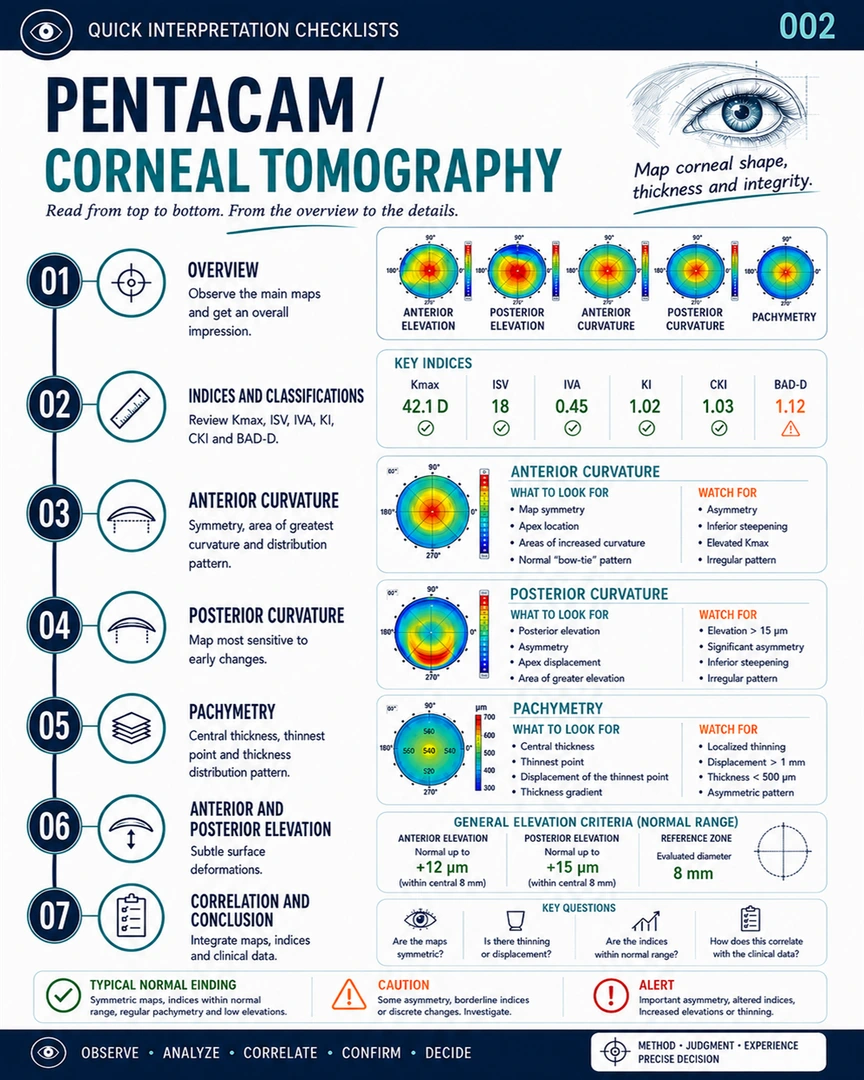
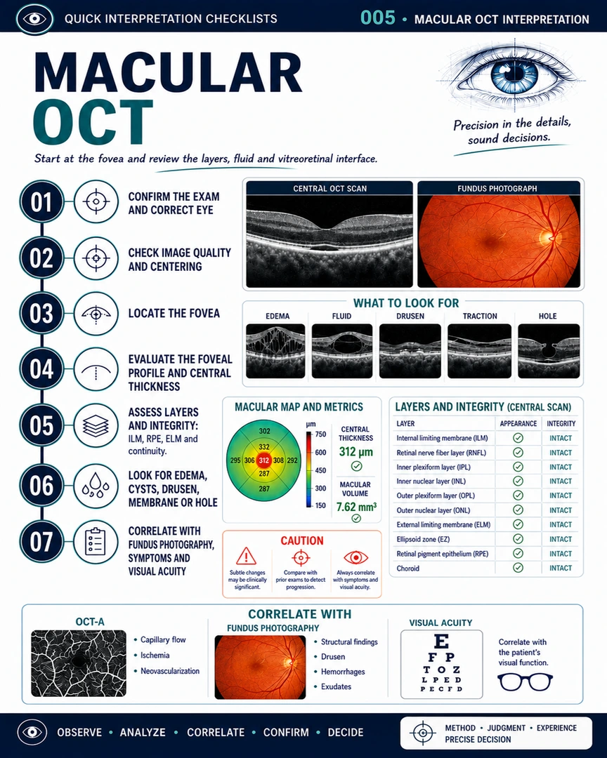
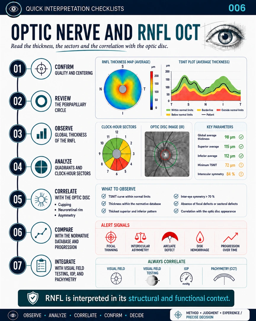
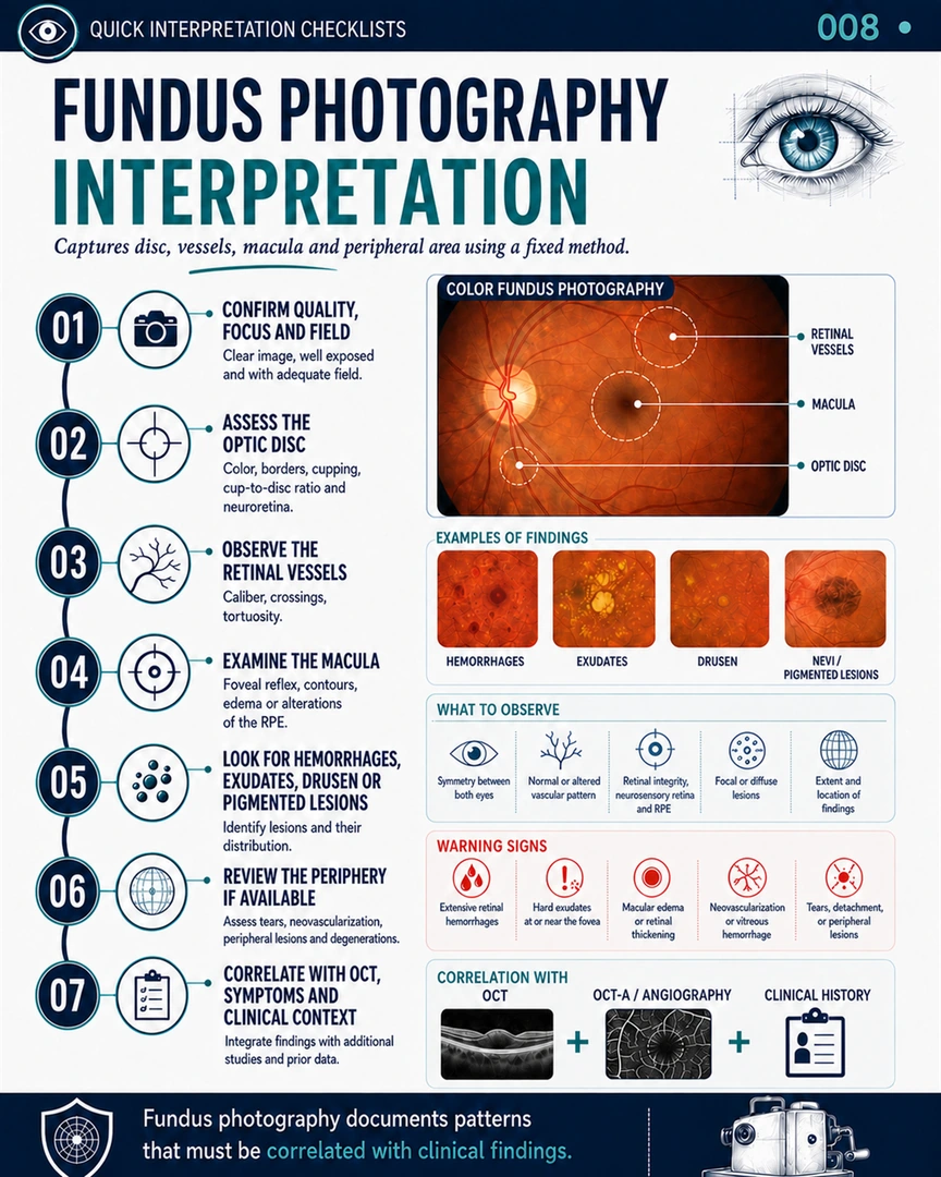
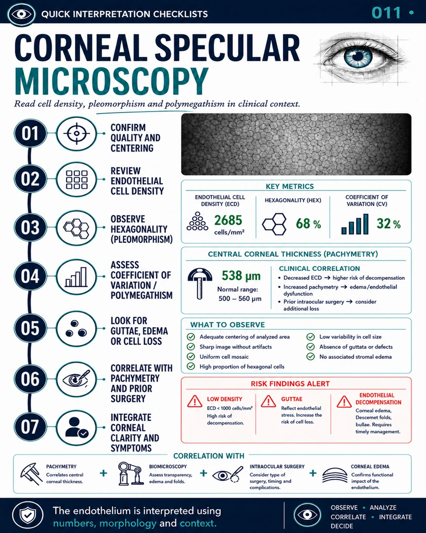
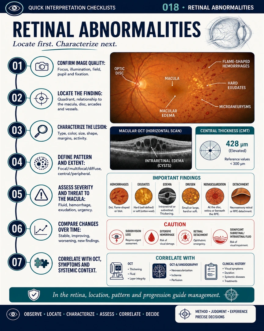
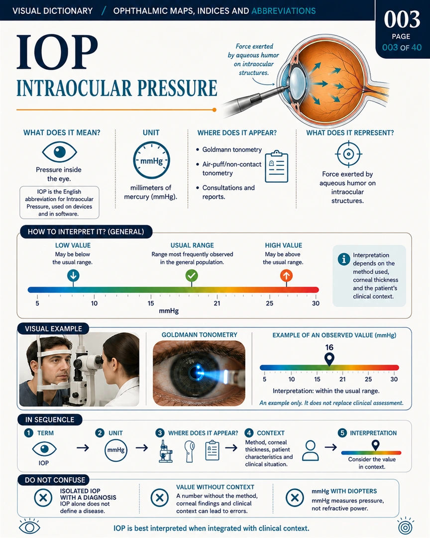
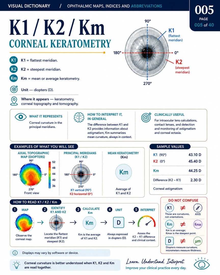
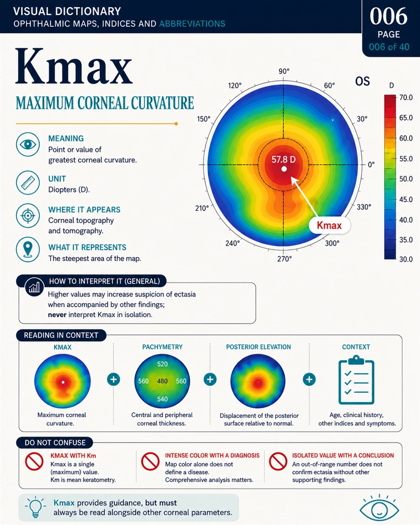
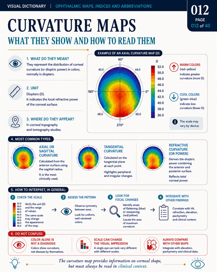
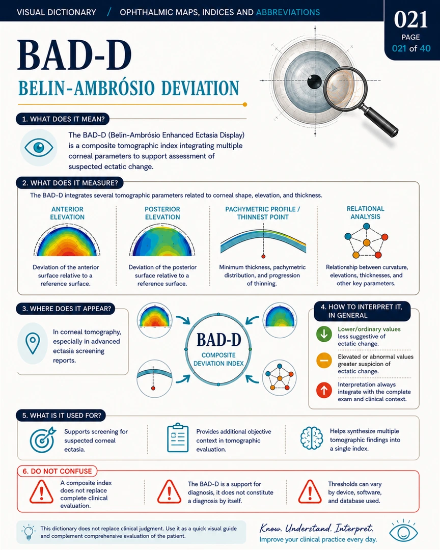
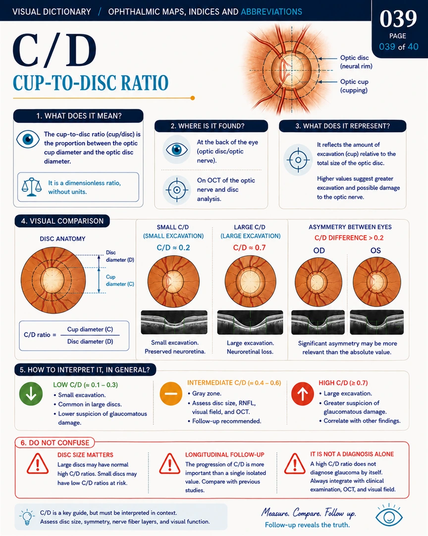
The system turns a screen full of colors, curves, indices, and images into a reading sequence: identify, validate, locate, interpret, compare, and integrate.
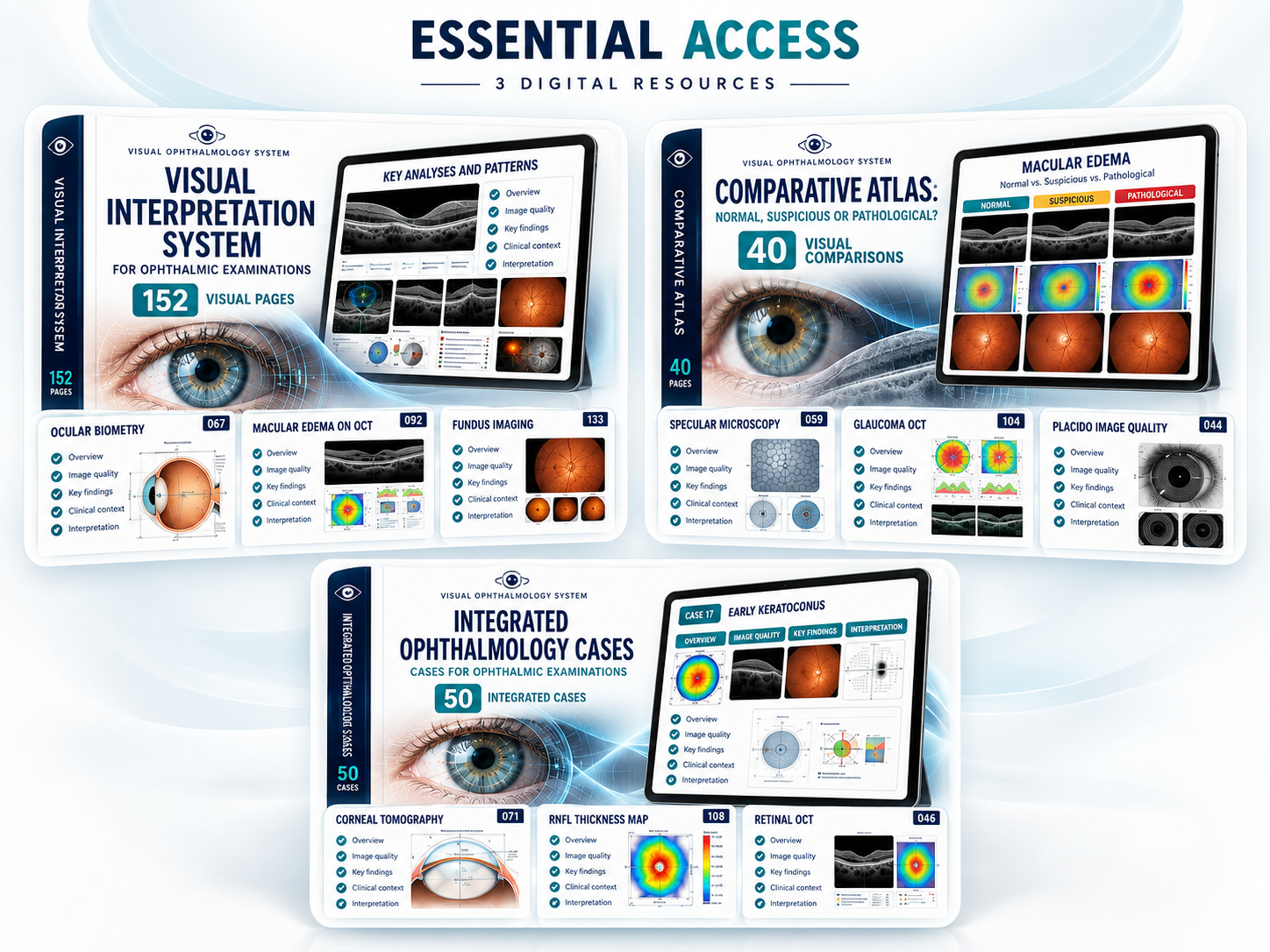
The core library combines the 152-page visual system, a 40-page comparative atlas, and 50 integrated cases to move from understanding the exam to applying the method.
Quick-reference tools to reinforce interpretation, spot artifacts, decode indices, and review high-yield patterns.
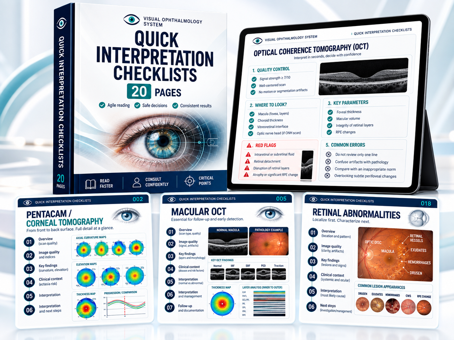
A clear sequence to keep the essentials in view: what to check, where to look, and what deserves attention.
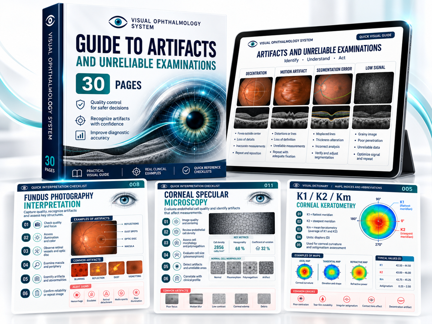
Not every image is reliable. Review acquisition errors, artifacts, and warning signs before you interpret.
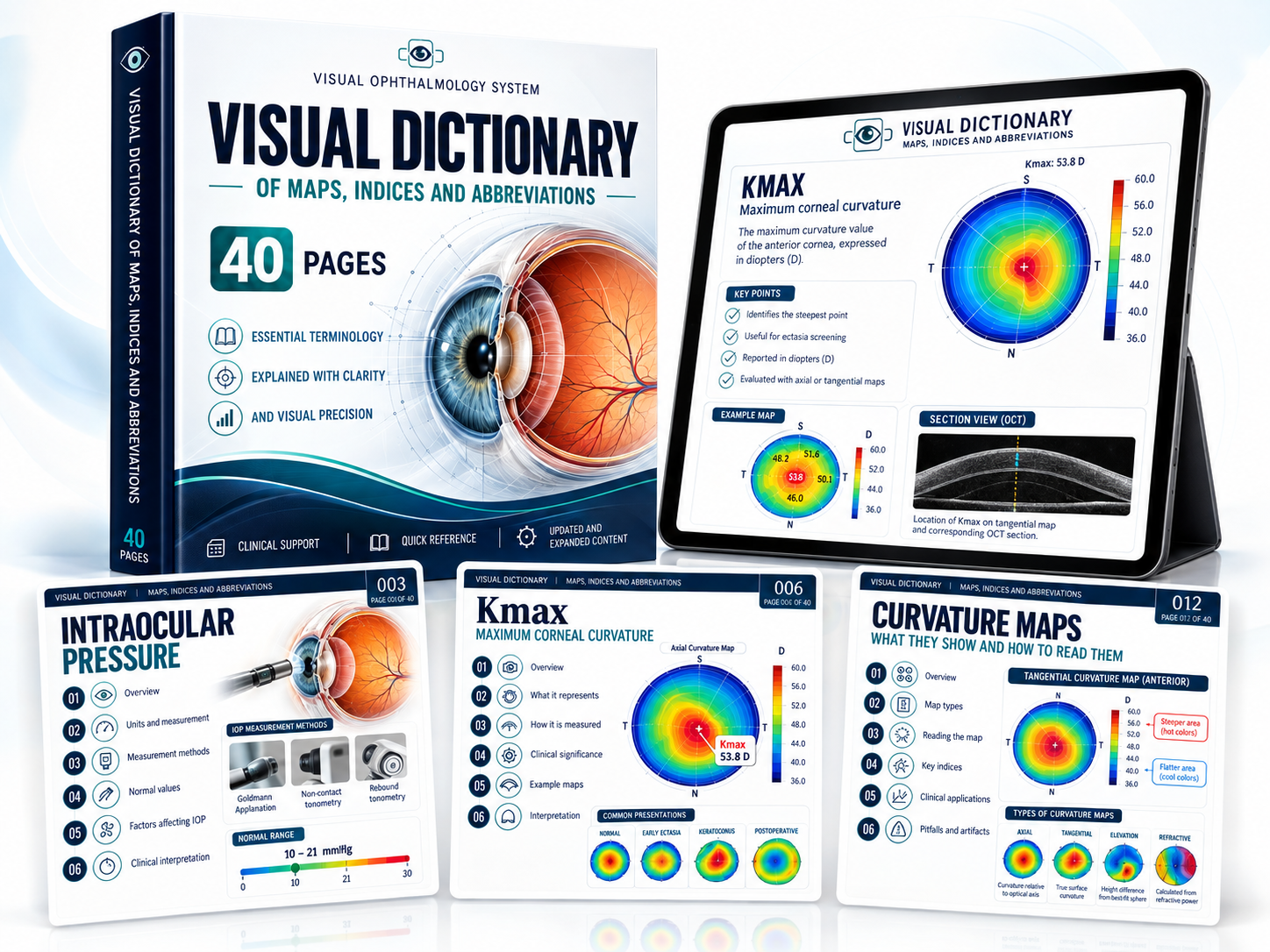
Keep key maps, indices, abbreviations, and parameters within reach while you study or review an exam.
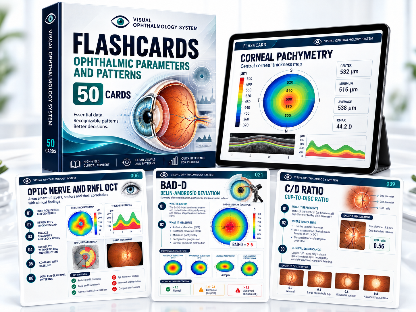
Use active recall to revisit ophthalmic parameters, patterns, and high-yield visual cues.
A fully digital product, with access provided after payment confirmation.
Complete your purchase using the payment methods available on the platform in your country.
Digital access to the materials is provided after payment confirmation.
View the PDFs on your phone, tablet, or computer, with no physical delivery to wait for.
Identify, validate, interpret, compare patterns, and apply what you learn to integrated cases.
The goal is not to memorize isolated numbers. It is to connect quality, maps, parameters, patterns, and context in a consistent sequence.
“Identify the examination in front of you and the information it can provide before you begin interpreting it.”“Distinguish a genuine variation from an artifact or an acquisition that does not meet reliability criteria.”“Read maps, colors, curves, and indices in a logical order, beyond memorizing isolated facts.”“Compare normal, suspicious, and pathological patterns to recognize differences that affect interpretation.”“Bring Pentacam, OCT, visual fields, and other examinations together into a more structured clinical picture.”“Quickly look up concepts, abbreviations, and parameters during study, review, or professional practice.”SWIPE TO SEE MORE
Start with the three core resources, or add all four bonus resources for US$ 7.00 more. Both options are one-time purchases with digital access.
Three core resources to build a visual approach to ophthalmic examination interpretation.
Study, compare, practice, and review with the entire Visual Ophthalmology library.
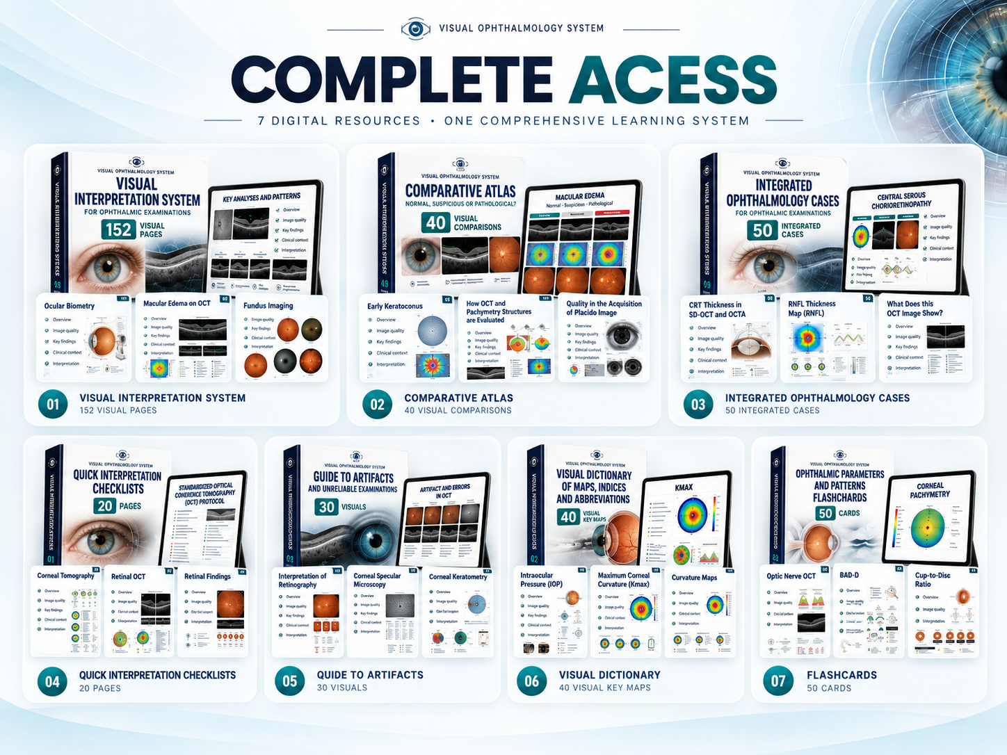
380+ pages, 50 cases, checklists, a visual dictionary, and flashcards
What you need to know about the format, content, and the two access options.
This is a fully digital product. After payment confirmation, access is provided through the platform's delivery process.
This edition is in English, for students and professionals in English-speaking markets worldwide.
It covers Pentacam, corneal tomography and topography, pachymetry, specular microscopy, biometry, aberrometry, retinal OCT, optic nerve OCT and glaucoma, visual field testing, fundus photography, and other key topics.
Essential Library includes the 3 core resources. Complete Library includes those same 3 resources plus the 4 specialized bonus resources shown on this page.
Yes. The visual sequence starts with identifying the examination and checking its quality, then progresses to pattern recognition and integrated cases.
Yes. You can view the digital materials on your phone, tablet, or computer.